2023. 3. 6. 19:25ㆍ카테고리 없음
안녕하세요! 헬씨부입니다~

오늘은 ' 한센병[ leprosy ] '에 대해 알아보고 정리하겠습니다!
한센병
[ leprosy ]
-요약-
나균에 의한 감염증으로 나균이 피부, 말초 신경계, 상부 기도를 침범하여 병적인 변화를 일으키는 만성 전염성 질환
정의
나균에 의해 감염되는 만성 전염성 질환을 한센병이라고 합니다. 6세기에 처음 발견된 병이며, 현재는 전 세계적으로 24개국을 제외한 나머지 지역에서 연간 1만 명당 1건 미만으로 발생하는 드문 질환입니다. 나균은 피부, 말초신경계, 상기도의 점막을 침범하여 조직을 변형시키게 됩니다.
한센병이라는 명칭은 노르웨이 의사 한센에 의해 나환자의 결절에서 나균이 처음 발견된 것에서 유래하였습니다. 한의학에서는 가라, 풍병, 대풍라 등으로 불렸고, 과거에는 문둥병 또는 천형병으로 불렸습니다. 현재는 일부 학술적 분야에서는 나병으로 하되, 사회적 분야에서는 한센병으로 통칭하고 있습니다. 왜냐하면 나병이란 용어가 편견과 차별적인 의미가 내포되어 사용되었기 때문입니다.
* 발병위치 : 피부, 말초신경계, 상기도
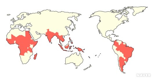
전 세계 한센병의 분포
원인
나균이 원인 병원체입니다. 이는 가족 내에서 장기간의 긴밀한 접촉으로 인해 전파되는 것으로 알려졌으나 정확한 감염 경로는 아직 밝혀지지 않았으며, 상기도나 상처가 있는 피부를 통해 나균이 침입하는 것으로 추측되고 있습니다.
증상
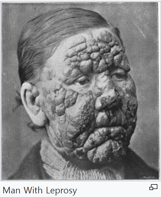
잠복기간은 9개월~20년으로 다양합니다. 한센병은 피부에 나타나는 병적인 변화의 종류에 따라 크게 나종한센병(lepromatous leprosy; 나종나)과 결핵한센병(tuberculoid leprosy; 유결핵형, 유결핵나)의 두 가지 형태로 나눌 수 있고, 두 종류의 중간 단계에 해당하는 다양한 양상이 관찰됩니다.
나종한센병의 잠복기는 결핵한센병의 잠복기의 두 배 정도로 깁니다. 나종한센병의 경우 전신의 피부에 양쪽 대칭적으로 결절(지름 5mm 이상의 발진)이나 구진(좁쌀 크기에서 완두콩 크기까지의 지름 5mm 이하인 발진) 등의 병변이 넓게 나타납니다. 나균이 코 점막에 침범하면 딱지가 생기고, 코막힘, 출혈 등을 일으키며, 눈에 침범하면 홍채염이나 각막염을 일으킵니다.
결핵한센병의 경우에는 한 개 이상의 경계가 뚜렷한 피부염이 신체에 비대칭적으로 퍼져 나타나고, 증상이 나타난 피부 부위는 무감각 또는 과다 감각 상태가 됩니다. 결핵한센병은 특히 말초신경으로의 나균 침범이 심하게 일어납니다.
경계형한센병(borderline leprosy)은 치료하면 결핵한센병으로, 치료하지 않으면 나종한센병으로 변합니다. 질병의 초기 단계에 나타나는 부정형한센병(indeterminate leprosy, 부정한센병)으로 인해 경계가 불분명한 저색소 반상(반점)이 나타나고, 이를 치료하지 않으면 나종한센병, 결핵한센병, 경계형한센병으로 진행됩니다. 한센병이 경과되면서 한센병 반응(leprosy reaction)이라는 상태가 나타나는데 이는 질병의 악화를 의미합니다. 나종한센병에서는 한센병성 결절성 홍반(ENL:erythema nodosum leprosum) 반응이, 경계형한센병에서는 역전반응(reversal reaction)이 나타납니다.
*발병위치 : 피부, 말초신경계, 상기도
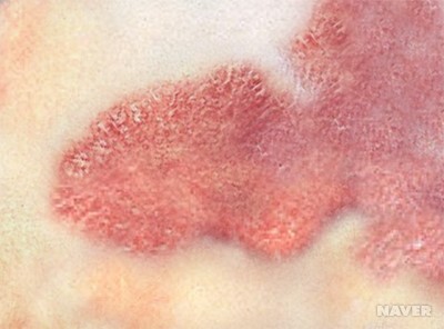
나종한센병의 피부병변
진단/검사
피부에 나타나는 변화를 확인하고, 말초신경이 침범되었을 때 나타나는 증상인 과다 감각, 감각소실, 마비, 근육 위축, 양측 말초 신경의 비대나 압통 등의 확인을 통해 진단합니다. 병적인 변화가 나타나는 피부나 신경 조직에 특이 염색을 시행하여 나균을 확인할 수 있습니다. 나종한센병의 진단에는 피부조직 염색 검사가 유용하지만 결핵한센병의 경우에는 단위면적당 존재하는 나균의 수가 적기 때문에 유용하지 않습니다. 그 외 나균의 균체에 존재하는 페놀성 당지질에 의해 면역계가 자극되어 생성된 항페놀당지질-1 항체(anti-phenolic glycolipid-1 antibody)를 측정하거나 나균의 핵산을 검출하는 검사법이 있습니다.
치료
한센병의 치료에는 여러 종류의 항생제를 함께 쓰는 병합요법이 사용됩니다. 나균에 효과적인 항생제로는 댑손(dapsone), 리팜핀(rifampin), 클로파지민(clofazimine) 등이 있습니다.
경과/합병증
치료하지 않으면 신경계의 합병증으로 인해 사지의 무감각과 근육의 병적인 증상이 발생합니다. 촉감, 통각, 온도 감각이 소실되고 위치감각과 진동감각도 없어집니다. 나균에 가장 흔히 침범되는 신경 부위는 팔꿈치이며 4번째와 5번째 손가락이 갈퀴처럼 변형됩니다. 손목 처짐이나 발목 처짐이 발생할 수 있습니다. 손가락과 발가락에 감각이 소실된 상태에서 지속적으로 외상을 입고 이로 인해 이차 감염이 발생하면 손가락과 발가락의 말단 부위가 떨어져 나가기도 합니다.
코점막에 나균이 침범하면 코피가 나거나 코막힘 증상이 나타나고, 코연골의 만성적인 염증으로 인해 연골이 변형되어 안장코(saddle nose)가 되거나 무후각증이 발생하기도 합니다. 눈이 침범되면 안구가 돌출되거나 눈이 감기지 않게 되고 각막궤양이나 백내장, 녹내장 등이 발생하여 실명할 수도 있습니다. 나균이 고환염을 일으킬 경우 무정자증이 되어 불임이 될 수도 있습니다.
문제
A 29-year-old man presents to the clinic complaining of fatigue and loss of sensation in his lower legs. He denies any history of trauma or chronic disease but states that he spends a lot of time outside and often encounters wild animals. On examination, he has multiple pale lesions over the skin of his face and back, decreased sensation of fine touch and vibration from mid-calf to foot bilaterally, and ulcerative lesions to the soles of both feet which are not painful to palpation. What is the morphology of the organism that is causing this man’s symptoms?
|
|
Explanation:
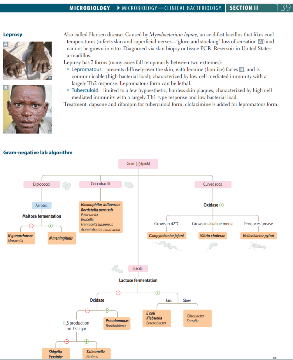
Correct Answer B: An acid-fast, intracellular bacillus: Leprosy (Hansen’s disease) is caused by the bacteria Mycobacterium leprae. It thrives in cooler temperatures, which is why it can be found infecting the skin and peripheral nerves. This man presents with hypopigmented skin lesions, ‘stocking-glove’ neuropathy, and painless plantar ulcers that are typical of lepromatous Hansen’s disease. Progression involves blindness and leonine facies. In the United States, Hansen’s disease is most often caused by contact with armadillos, an animal reservoir of the bacteria.
Option A: A spirochete transmitted via tick: Borrelia burgdorferi is a double-membraned spirochete that causes Lyme disease. This bacteria is transmitted via the Ixodes scapularis, or deer tick, which is found primarily in the northeastern United States. Lyme disease is a protracted illness characterized by a bullseye rash, chronic fatigue, transient arthritis, and eventually encephalopathy and carditis. The bullseye rash, also known as erythema migrans, is very identifiable. This man’s symptoms are not characteristic of Lyme disease, making this an unlikely diagnosis.
Option C: Gram-positive, branching anaerobe: Actinomyces species have a characteristic appearance as a branching, filamentous rod that resembles a branching fungus. It is a normal flora of the oral cavity that, in response to orofacial trauma, may cause an abscess. Sinus tracts typically form, and the drainage contains yellow sulfur granules. This man’s presentation is not consistent with infection with this bacteria.
Option D: Maltose-fermenting gram-negative diplococci: Neisseria meningitidis is a gram-negative diplococcus that can be identified based on its ability to ferment maltose on agar. N. meningitidis is the most common cause of meningitis in young adults, especially those in close quarters like college students and military recruits. Onset is sudden, and the illness is progressive, leading to shock. Waterhouse-Friderichsen syndrome, the hemorrhagic destruction of both adrenal glands, is a possible complication. The rash caused by N. meningitidis is initially petechial, progressing to purpura.
Option E: Reactivation of latent viral infection: Herpes zoster (shingles) is a painful rash that appears in a dermatomal pattern due to the reactivation of varicella-zoster virus (VZV). Initial infection with VZV causes chickenpox, a disease characterized by fever and a diffuse pruritic rash. Once the illness resolves, the virus stays latent in neural ganglia and can become reactivated later in life. Shingles does not cause systemic symptoms or loss of sensation. Treatment is with antivirals in order to reduce the risk of developing post-herpetic neuralgia, defined as pain that persists in the area of the rash for more than four months.
Learning objective: Hansen’s disease is characterized by cutaneous and neurologic findings. It is caused by Mycobacterium leprae, an acid-fast intracellular bacillus.
|
|
Book References:
First Aid for the USMLE Step 1 (2022, 32nd ed): 139
First Aid for the USMLE Step 1 (2021, 31st ed): 141
First Aid for the USMLE Step 1 (2020, 30th ed): 141
First Aid for the USMLE Step 1 (2019, 29th ed): 141
First Aid for the USMLE Step 1 (2018, 28th ed): 141
First Aid for the USMLE Step 1 (2017, 27th ed): 137
|
참고자료
오늘은 여기까지 정리하겠습니다!
감사합니다!