2023. 2. 26. 14:36ㆍ카테고리 없음
안녕하세요! 건강정보통통 입니다~

오늘은 ' 흔들린아이증후군[ Shaken baby syndrome ] '에 대해 알아보고 정리하겠습니다!
흔들린아이증후군
[ Shaken baby syndrome ]
-요약 -
2세 이하의 유아를 심하게 흔들어서 생기는 질환

정의
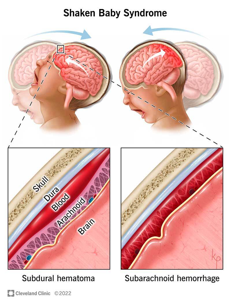
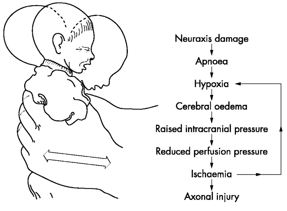
영유아에서 양육자가 고의로 아이를 강하게 흔들어 생기는 두부손상의 형태이다. 주로는 2세 이하의 소아에서 많이 발생한다. 아이가 울음을 그치지 않으면서 잘 달래지지 않는 상황에서 분노의 표현으로 아이를 흔들어 대면서 발생한다. 가정 내에 폭력상황이 빈번이 발생하거나 양육자가 약물중독에 빠져 있는 경우가 있다. 아이를 세게 앞 뒤로 흔들면 두부의 급가속/감속 충격이 뇌에 전달되면서 지주막하출혈, 경막하출혈, 뇌실질손상이 올수 있다.
원인
영유아의 목 근육의 근력은 약한데 비해 머리크기는 상대적으로 커서 앞 뒤로 아이를 강하게 흔들게 되면 심각한 두부 손상을 초래할 수 있다.
증상
영유아에서 경막하출혈, 지주막출혈 등의 심한 두부손상이 발견되었으나 원인을 설명할 수 있는 외부요인이 없는 경우에 꼭 의심하여야 한다. 증상은 사건 발생 직후부터 나타날 수 있으나 4-6시간 후에 가장 심각해진다. 의식과 반응이 떨어지고 심하면 혼수상태에 빠질 수 있다. 구토가 반복되고 호흡이 불규칙해 질 수 있다. 경련/발작이 동반된다.
진단/검사
응급의학과 혹은 소아청소년과 전문의의 검진이 필요하다.
뇌영상(CT 혹은 MRI)검사와 망막검사(망막출혈)가 필요하다.
치료
뇌출혈 및 뇌부종에 대해서 신속한 진단과 치료가 필요하다. 내과적인 처치로 호전이 없을 경우 혈종제거 및 감압술 같은 뇌수술이 필요할 수 있다.
경과/합병증
손상의 범위가 광범위하고 치료가 늦어지는 경우 사망하게 될 수 있다.
생존하더라도 뇌성마비, 지적장애, 뇌전증 등의 합병증이 발생하는 빈도가 높다.
예방방법
아이를 처음 양육하는 부모에게 적절한 양육방법 및 아이가 울고 달래지지 않을 때의 대처법에 대한 교육과 상담이 필요하다.
문제
A 2-month-old boy is brought to the emergency room by his mother for a 3-hour history of decreased responsiveness. She reports that she left the baby with a new nanny this morning and he was behaving normally. When she got home in the afternoon, he seemed lethargic and would not breastfeed as usual. At birth, the child had an Apgar score of 8/9 and weighed 2.8 kg (6.1 lb). Growth has been in the 90th percentile and the patient has been meeting all developmental milestones. There is no significant past medical history and vaccinations are up-to-date. On physical exam, the patient does not seem arousable. Ophthalmologic evaluation reveals retinal hemorrhages. Which of the following findings would most likely be expected on a noncontrast CT scan of the head?
|
Explanation:
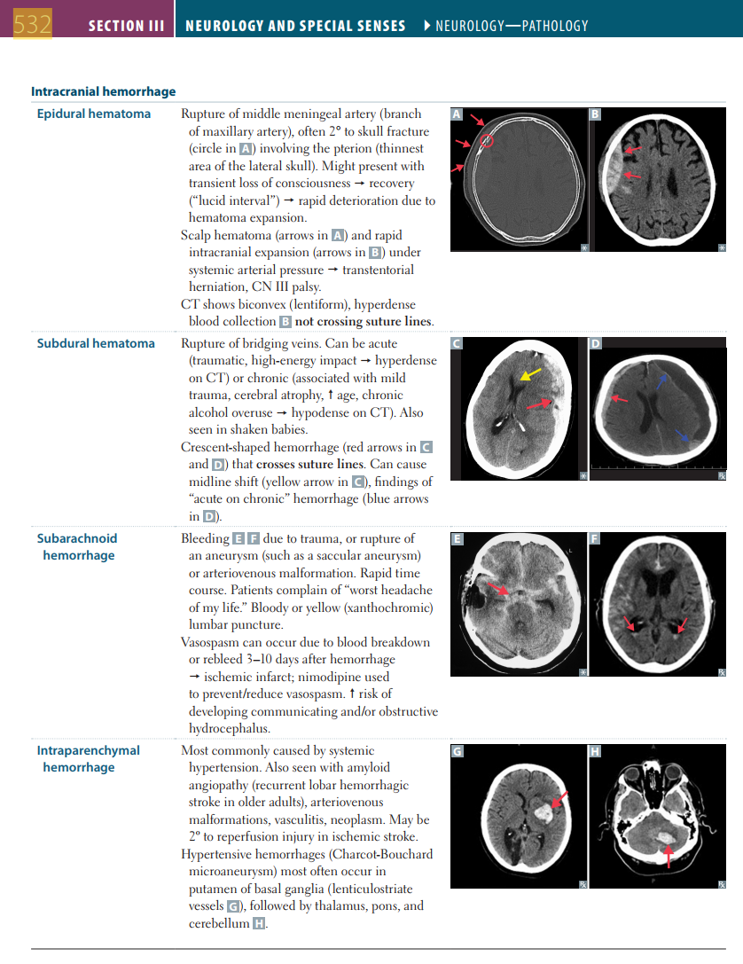
Correct answer A: A subdural hematoma occurs due to the rupture of bridging veins and can be acute or chronic. An acute subdural hematoma is seen in "shaken baby syndrome," which may occur in children who have been injured by forceful shaking or trauma. Chronic subdural hematomas are associated with cerebral atrophy, old age, and alcoholism. Typical CT findings include a crescent-shaped hyperdensity that crosses the suture line, but not the midline.
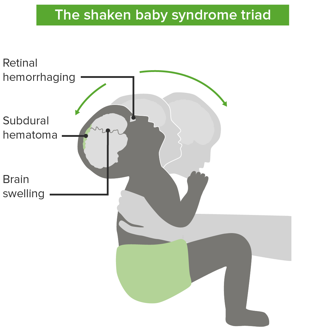
Image: Diagram showing typical injuries of the "shaken baby syndrome." By Lecturio
Option B: An epidural hematoma typically results from the rupture of the middle meningeal artery due to a lateral skull fracture. Clinically, patients classically present with a lucid interval and subsequently become acutely unresponsive. CT shows a biconvex lens-shaped, hyperdense collection of blood that does not cross suture lines.
Option C: A subarachnoid hemorrhage occurs due to the rupture of a saccular aneurysm (i.e. berry aneurysm) or rupture of an arteriovenous malformation. It is often described by patients as the worst headache of their life. Lumbar puncture reveals blood in the cerebrospinal fluid. Complications include vasospasm and hydrocephalus.
Option D: Alzheimer disease is the most common cause of dementia in the elderly. It is characterized by amyloid deposition in the brain (i.e. senile plaques) and accumulation of hyperphosphorylated insoluble tau protein in the cytoplasm (i.e. neurofibrillary tangles). Neuroimaging typically shows widespread cortical atrophy with narrowing of the gyri and widening of the sulci.
Option E: Vascular dementia is the second most common cause of dementia in the elderly and is due to a progressive impairment that results from multiple small ischemic infarcts. Neuroimaging shows multiple cortical and subcortical infarcts.
|
Intracranial hemorrhages
|
||
|
|
Causes & CT findings
|
Images
|
|
Epidural hematoma
|
Cause: Rupture of the middle meningeal artery due to a lateral skull fracture.
Patients usually present with a lucid interval.
CT findings: Biconvex or lentiform hyperdense blood collection
|
Image: Postoperative CT scan. By Eftekhar et al, License: Public Domain
|
|
Subdural hematoma
|
Causes: Rupture of bridging veins due to trauma ("shaken baby syndrome") if acute. If chronic, it may be due to alcoholism, old age, or cerebral atrophy.
CT findings: Crescent-shaped hemorrhage that crosses suture lines, with or without a midline shift
|
Image: Subdural hematoma. By Chye et al, License: Public Domain
|
|
Subarachnoid hemorrhage
|
Causes: Saccular or berry aneurysm; arteriovenous malformations.
Presents with a severe headache. Risk of communicating or obstructive hydrocephalus.
CT findings: Blood in the basal cisterns
|
Image: Subarachnoid hemorrhage seen on CT scan. By Hellerhoff, License: CC BY-SA 3.0
|
|
Intraventricular hemorrhage
|
Causes: Complication of prematurity (usually within the first 5 days), bulging fontanelle, hypotension, seizures, irregular respiration, coma.
Bleeding from the germinal matrix (vascularized layer in the subventricular zone that usually regresses).
CT findings: Blood in the ventricles
|
Image: Intracerebral hemorrhage. By Glitzy queen00, License: Public Domain
|
|
Intraparenchymal hemorrhage
|
Causes: Hypertension, amyloid angiopathy, vasculitis, neoplasm, reperfusion injury
|
Typically in basal ganglia and internal capsule
|
Learning objective: Subdural hematoma occurs due to the rupture of bridging veins and can be acute or chronic. An acute subdural hematoma is seen in "shaken baby syndrome," a form of child abuse or trauma, while a chronic subdural hematoma is associated with cerebral atrophy, old age, alcoholism. On CT, a crescent-shaped hemorrhage is seen that crosses the suture line and often causes a midline shift.
|
Related Videos:
|
|
Book References:
First Aid for the USMLE Step 1 (2022, 32nd ed): 532
First Aid for the USMLE Step 1 (2021, 31st ed): 531
First Aid for the USMLE Step 1 (2020, 30th ed): 513, 556, 706
First Aid for the USMLE Step 1 (2019, 29th ed): 501
First Aid for the USMLE Step 1 (2018, 28th ed): 497, 540
First Aid for the USMLE Step 1 (2017, 27th ed): 483, 526
|
참고자료
https://terms.naver.com/entry.naver?docId=6225906&cid=51007&categoryId=51007
오늘은 여기까지 정리하겠습니다!
감사합니다!