2022. 12. 8. 16:51ㆍ카테고리 없음
안녕하세요! 헬씨부 입니다~
오늘은 ' 흡수장애 증후군 [Malabsorption Syndrome] '에 대해 알아보고 정리하겠습니다!

흡수장애 증후군
[Malabsorption Syndrome]

정의
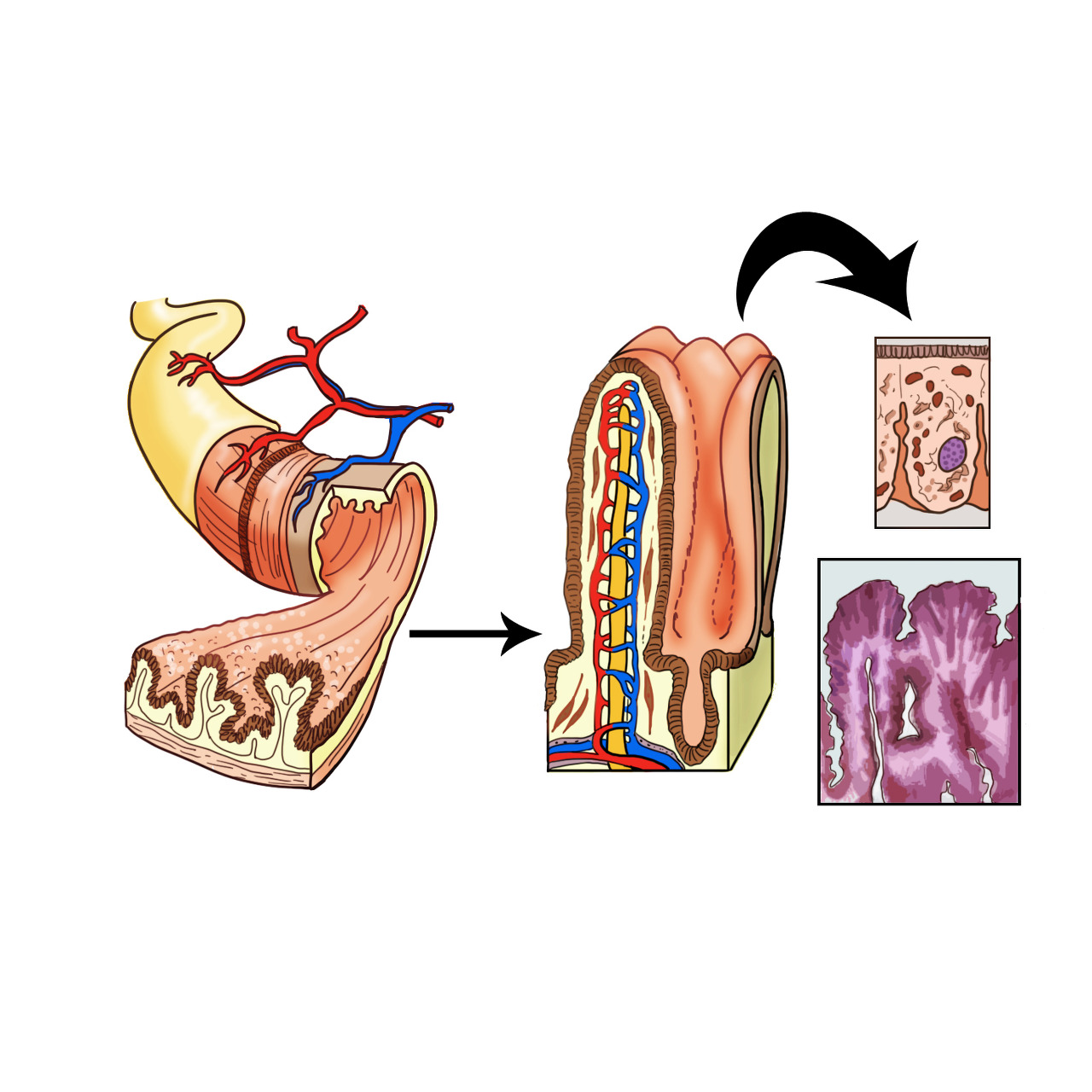
흡수장애 증후군은 먹은 음식물을 소화하여 흡수하는 과정 중 어느 한 부분이라도 문제가 있어 발생하는 증상을 말하는 질환입니다. 이는 소화 효소에 의해 분해가 잘 이루어지지 않는 흡수장애와 분해된 영양소를 장세포 내로 흡수하지 못하는 흡수장애로 나눌 수 있습니다.
원인
흡수장애 증후군은 장관 내에 이상이 발생한 경우, 담관이 막혀서 담즙이 나오지 못하거나 만성 췌장염으로 인하여 췌장 소화액 분비가 부족한 경우, 소장 내에 세균이 증식하여서 세균이 소화 효소를 분해하는 경우 등이 원인이 됩니다.
흡수면 부족에 의한 흡수장애는 다른 질환으로 인하여 소장을 절제하여 소장이 100cm 이내만 남아있는 짧은 소장(단장)에서 나타날 수 있습니다. 소장의 마지막 흡수를 담당하는 소장의 융모가 파괴되는 경우에도 흡수 면적이 줄어 들어서 흡수가 잘 되지 않습니다.
이외에 원인으로 감염, 장관 내의 가수 분해나 가용성의 감소, 점막 세포의 이상, 림프관의 폐쇄, 방사선 치료 등이 있습니다.
증상
흡수장애는 성장과 발달에 영향을 미치거나 특정 질환을 유발할 수 있습니다. 식품으로부터 영양소를 흡수하지 못해 설사, 더부룩함, 복통, 다량의 변, 근육 위축 등이 나타나게 됩니다.
소장 내 세균이 과다 증식해서 담즙 대사에 장애가 오면 지방 흡수에 장애가 발생하여 지방변을 보게 됩니다. 빈혈(철분이나 엽산, 비타민 B12)이나 비타민 K의 부족으로 출혈이 발생할 수 있고, 비타민 A, B1 결핍으로 내분비 질환이 발생거나 말초 신경증이 발생하기도 합니다. 단백질 흡수장애로 부종이 생기기도 합니다. 피부에는 피부염과 각질층이 많이 생기는 현상이 뚜렷해지기도 하며, 골다공증과 강직 경련이 일어날 수 있습니다.

진단/검사
흡수장애 증후군 진단은 혈액 검사를 통해 전해질 수치, 콜레스테롤 수치, 알부민 수치, 프로트롬빈 시간 측정을 진행함으로써 진단합니다. 대변 검사를 통해 지방변을 보는지를 검사합니다. 소화관 x-ray 검사, 소장의 점막 생검, Xylose 흡수, secretin-test 등의 검사를 시행하여 흡수장애 증후군을 진단합니다.
치료
흡수장애 증후군 치료는 우선 정맥 주사를 통하여 고칼로리의 영양제를 주입하여 영양 보충합니다. 이후 흡수장애의 원인이 되는 질환을 치료합니다.
경과/합병증
흡수장애 증후군을 겪으면 각종 비타민 부족으로 야맹증이나 피하 출혈이 일어날 수 있습니다. 피부가 닭살처럼 변모하는 경우도 있고, 빈혈이 심해서 창백해지기도 합니다. 흡수장애 증후군으로 인해 골연화증이 생기면, 뼈가 휘고 잘 부러지게 되며 사망에 이르기도 합니다.
문제
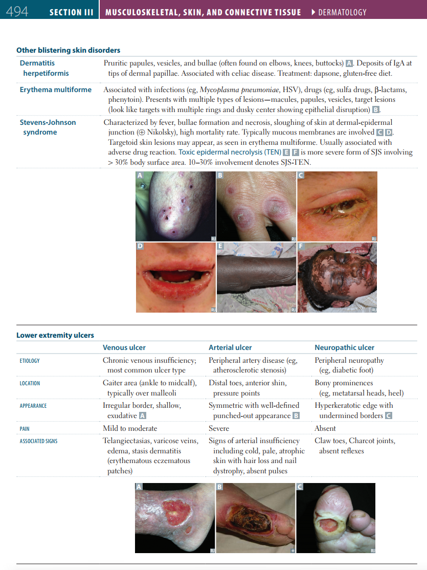
A 45-year-old man presents to the office for evaluation of pruritic skin lesions on his elbows and knees. He says that they have been present for one month and that he has been using over-the-counter ointments to treat them, but those have not helped. He has not seen a healthcare provider for many years. He has no known allergies. Blood pressure is 140/80 mm Hg, pulse is 82/min, respiratory rate is 18/min, and temperature is 37.2°C (98.9°F). On examination, clustered vesicular lesions are noted on both elbows and knees. Cardiovascular and pulmonary exams are unremarkable. Which of the following would most likely be associated with this man’s condition?
|
|
Explanation:
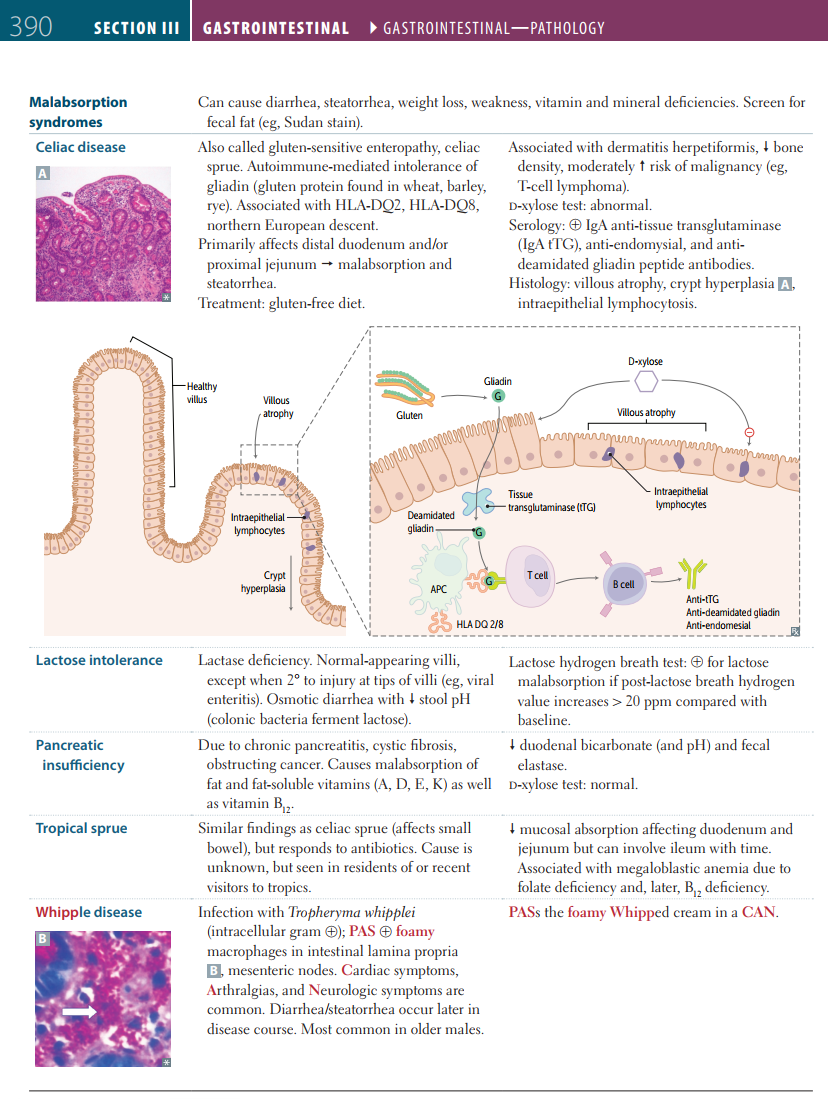
Correct Answer A: Malabsorption. The man's rash is dermatitis herpetiformis (DH), which is associated with celiac disease. DH classically appears on extensor surfaces and resembles the vesicles of herpes simplex infections. The mechanism underlying celiac disease involves IgA antibodies against gliadin and/or transglutaminase complexes in the intestine. It is believed that these antibodies cross-react with transglutaminase in the skin. Celiac disease is known to cause malabsorption, which occurs due to inflammation of the intestinal lining after the consumption of gluten. Therefore, a diet restricting gluten is recommended and is proven to decrease gastrointestinal and dermatologic symptoms. Option B: Transmural inflammation of the colon and non-caseating granulomas are complications of Crohn’s disease, but they are not seen in celiac disease. Pyoderma gangrenosum is associated with Crohn's disease and appears as an ulcerative lesion, typically of the lower extremity, and is not similar to this man's rash. Option C: A double bubble on X-ray usually occurs in duodenal atresia in infants and represents dilatation of the proximal duodenum. Dermatologic findings are not usually found. Options D: Erythema nodosum is associated with various infections, cancers, and inflammatory bowel diseases, which include Crohn’s disease and ulcerative colitis. Erythema nodosum consists of painful nodules that arise in the pretibial area and may limit movement. It is not similar in appearance or location to this man's rash. Image: A person with erythema nodosum presenting with characteristic nodules on the shins. By James Heilman, MD, License: CC BY-SA 3.0 Option E: Acanthosis nigricans, which is velvety hyperpigmentation of the flexural skin, is usually seen in patients with diabetes types 1 and 2. Additionally, acanthosis nigricans may arise secondary to occult malignancies, such as gastric cancer. This man has none of those medical problems, and his rash is not similar to acanthosis nigricans. Image: Acanthosis nigricans on a diabetic patient. By Teelucksingh S, Jaimungal S, Pinto Pereira L, Seemungal T, Nayak S, Cardiovasc Diabetol (2012), License: CC BY 2.0 Learning objective: Dermatitis herpetiformis classically occurs secondary to celiac disease due to a shared pathologic mechanism. A gluten-free diet is recommended for both. |
|
Related Videos:
|
|
Book References:
First Aid for the USMLE Step 1 (2022, 32nd ed): 390, 494 First Aid for the USMLE Step 1 (2021, 31st ed): 391, 495 First Aid for the USMLE Step 1 (2020, 30th ed): 481 First Aid for the USMLE Step 1 (2019, 29th ed): 471 First Aid for the USMLE Step 1 (2018, 28th ed): 467 First Aid for the USMLE Step 1 (2017, 27th ed): 452 |
참고자료
앞선 의술 더 큰 사랑을 실천하는 서울아산병원 입니다
www.amc.seoul.kr
오늘은 여기까지 정리하겠습니다!
감사합니다!